Medial view of left knee region highlighting various fascial
$ 12.00
4.5(131)In stock
Download scientific diagram | Medial view of left knee region highlighting various fascial components surrounding the semitendinosus muscle. From the superficial to the deep aspect: the fascia lata, the paratenon and the epimysium from publication: Anatomical study of paratenons and fascia lata connections in the posteromedial knee region | Introduction In the last decade, fascia research increased significantly in various aspects such as anatomical and biomechanical features related to epimuscular force transmission. Methods The present anatomic study focuses on macroscopic observations of the potential | Fascia Lata, Hamstring muscles and Fascia | ResearchGate, the professional network for scientists.
A proposal for a new morphological classification of the popliteus
Marcel ROOZE, Université Libre de Bruxelles, Brussels, ULB, Laboratory of Anatomy, Biomechanics and Organogenesis
Anatomy of the Left Knee Medical Illustration Medivisuals
Benoit BEYER, Assoc. Prof., PT, MSc, PhD, Université Libre de Bruxelles, Brussels, ULB, Faculty of Motricity Sciences (FMS)
Medial view of right knee region highlighting gracilis (G) and
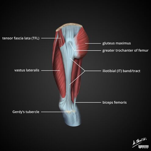
Iliotibial Tract - Physiopedia
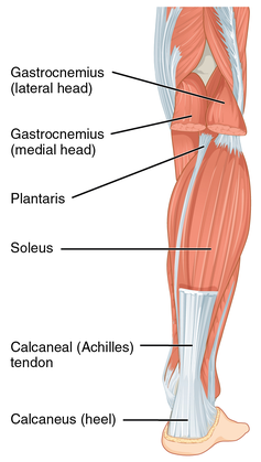
Calf Strain - Physiopedia
Fascial Manipulation Practical Part: Luigi Stecco, Carla Stecco
Perineal region: Anatomy, definition, diagram
Key Surgically Relevant Anatomy of the Medial and Lateral Aspects
1. Rana esculenta. (A) Pelvis (dorsal view); fascia removed on
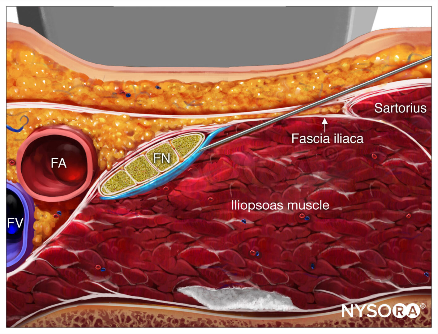
Ultrasound-Guided Femoral Nerve Block - NYSORA
Medial view of left knee region highlighting various fascial


/images/vimeo_thumbnails/258296721/UWQ0ucbOB1jHt8YDVJ8bQQ_overlay.jpg)