Ultra-wide-field fundus photographs and ultra-wide-field
$ 24.99
4.6(689)In stock
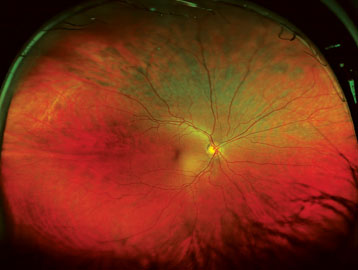
Download scientific diagram | Ultra-wide-field fundus photographs and ultra-wide-field fluorescein angiographic imaging of ocular toxocariasis. (A) A granuloma with mild vitreous opacity. (B) A tractional retinal fold with localized tractional retinal detachment. (C) Diffuse peripheral vascular leakage. (D) A prominent optic disc leakage. from publication: The Clinical Characteristics of Ocular Toxocariasis in Jeju Island Using Ultra-wide-field Fundus Photography | Toxocariasis, Ocular and Photography | ResearchGate, the professional network for scientists.
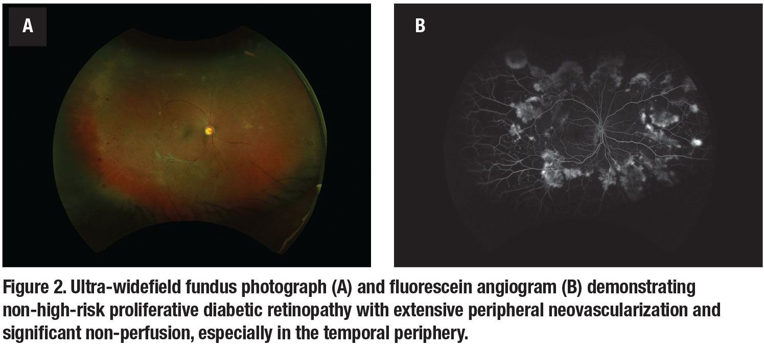
How ultra-widefield imaging is changing our view of DR
The Clinical Utility of Ultra-Wide-Field Imaging
Clinic-based ultra-wide field retinal imaging in a pediatric population, International Journal of Retina and Vitreous
Jong Young Lee's research works Jeju National University Hospital, Jeju City and other places
Ultra-wide-field fundus photographs and ultra-wide-field fluorescein
Clinic-based ultra-wide field retinal imaging in a pediatric population, International Journal of Retina and Vitreous
PDF) The Clinical Characteristics of Ocular Toxocariasis in Jeju Island Using Ultra-wide-field Fundus Photography
ZEISS CLARUS 500 Fundus Camera
Ultra-wide-field fundus photographs and ultra-wide-field fluorescein
Jong Young Lee's research works Jeju National University Hospital, Jeju City and other places
Eun Kyoung Lee's research works Dongguk University, Seoul and other places


:no_upscale()/cloudfront-us-east-1.images.arcpublishing.com/dmn/XK3ST37XKFEWDF5EONWQSRC5MA.jpg)