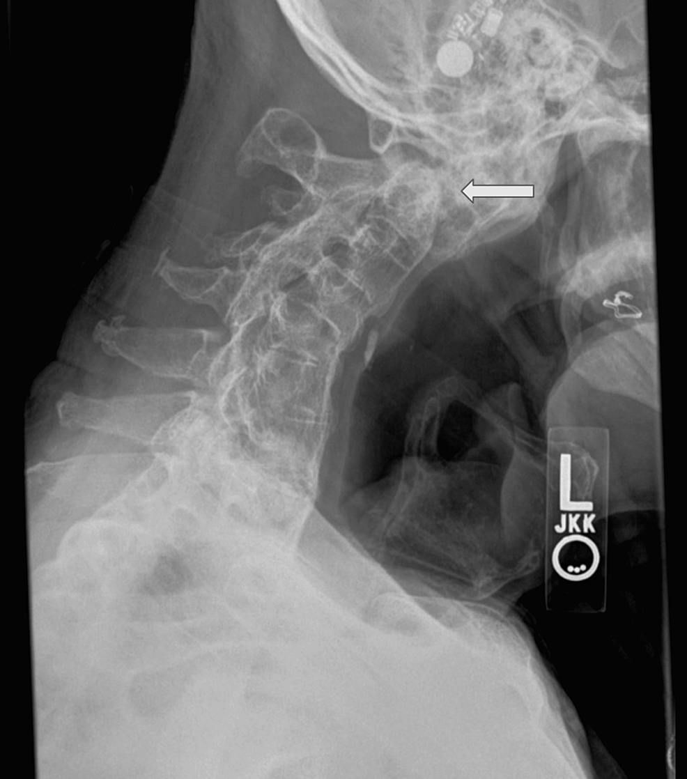
Lateral cervical spine showing C0-C3 fusion in reduced position after
Axial T1W (A) and T2W (B). Magnetic resonance imaging of the cervical
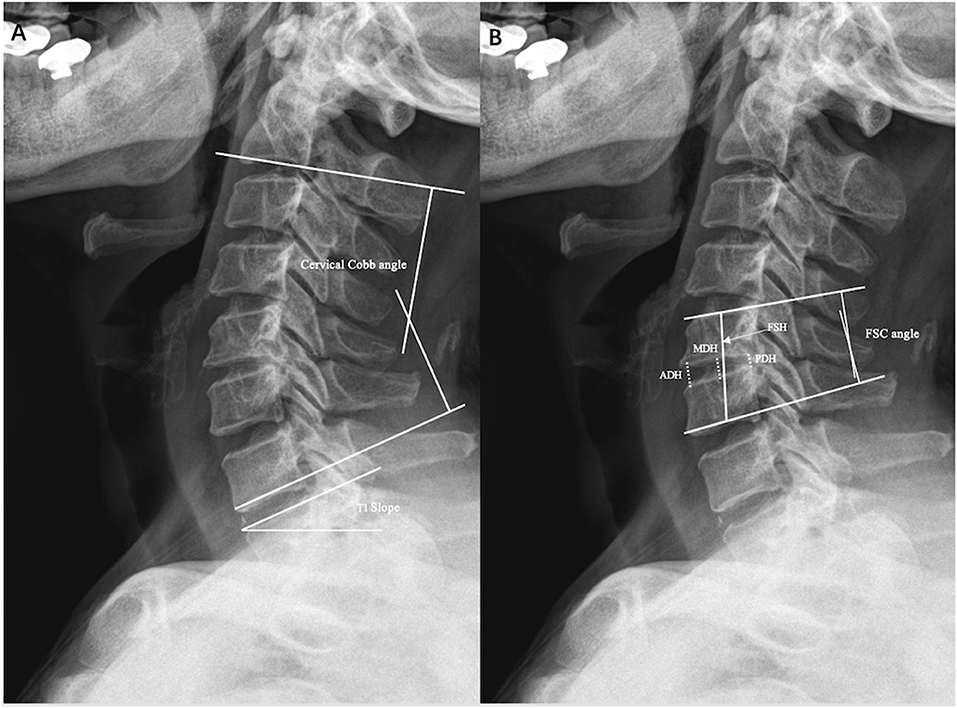
Frontiers Comparative Analysis of Cage Subsidence in Anterior Cervical Decompression and Fusion: Zero Profile Anchored Spacer (ROI-C) vs. Conventional Cage and Plate Construct
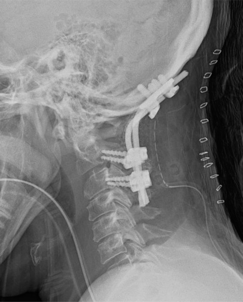
Instrumented reduction of a fixed C1–2 subluxation using occipital and C2/C3 fixation: A case report
Cureus, Posterior Cervical Fusion of Occiput-T3 for Unstable Complex Odontoid Fracture in an 80-Year-Old Male With C2-Sacrum Synostosis From Ankylosing Spondylitis: A Case Report