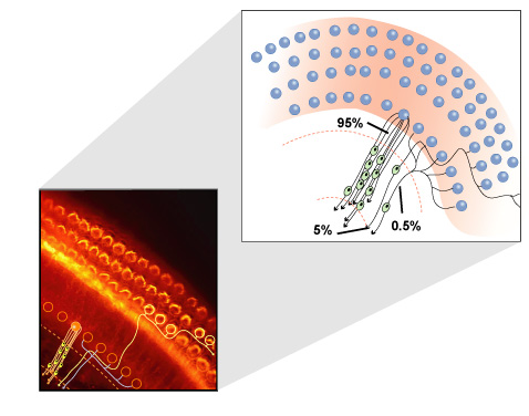
Innervation patterns of type I and type II auditory nerve fibers on
$ 10.00
4.5(96)In stock
Download scientific diagram | Innervation patterns of type I and type II auditory nerve fibers on inner and outer hair cells, respectively. Central and peripheral axons of type I cells are myelinated, whereas axons of type II neurons are unmyelinated. Peripheral terminals of type I and type II cells are unmyelinated within the organ of Corti, i.e. beyond the habenula perforata. from publication: Noise-induced and age-related hearing loss: New perspectives and potential therapies | The classic view of sensorineural hearing loss has been that the primary damage targets are hair cells and that auditory nerve loss is typically secondary to hair cell degeneration. Recent work has challenged that view. In noise-induced hearing loss, exposures causing only | Hair Cell, Hearing Loss and Neuro-Otology | ResearchGate, the professional network for scientists.
Auditory physiology of Inner Ear
Schematic drawing of the adult organ of Corti, the sensory epithelium
Animal models of hidden hearing loss: Does auditory-nerve-fiber loss cause real-world listening difficulties? - ScienceDirect
Auditory nerve definition of auditory nerve by Medical dictionary
Electron Microscopic Reconstruction of Neural Circuitry in the Cochlea - ScienceDirect
Auditory System: Structure and Function (Section 2, Chapter 12) Neuroscience Online: An Electronic Textbook for the Neurosciences
12 Diagram of the divergent innervation of five different cell types in
What are the examples of sensory and motor nerve fibres? - Quora
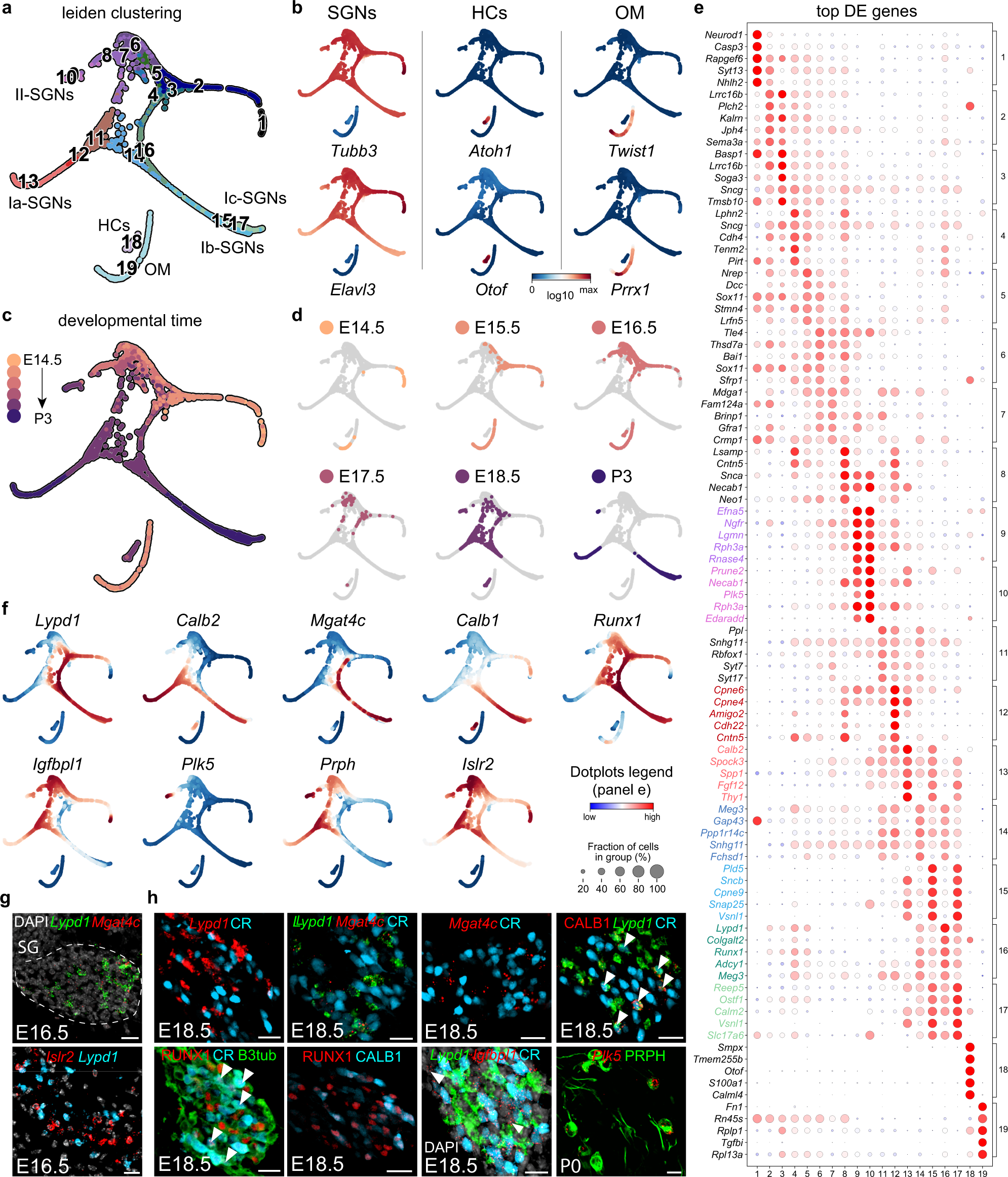
Single-cell RNA-sequencing analysis of the developing mouse inner ear identifies molecular logic of auditory neuron diversification
Figure 3 from Noise-induced and age-related hearing loss: new
Efferent Synaptic Transmission at the Vestibular Type II Hair Cell Synapse