Cellular fluorescence intensity and cell size as a function of
$ 19.50
4.9(92)In stock
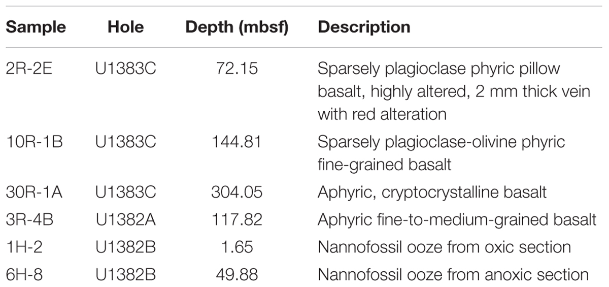
Download scientific diagram | | Cellular fluorescence intensity and cell size as a function of enrichment substrate on a subset of basalt enrichments (sample 30R-1A). The circle size indicates the average cellular area (mm 2 ) as measured by epi-fluorescence microscopy. The standard deviation for the cellular area of each sample is roughly 46% based on the following number of cells counts for each treatment: Host Rock (n = 28); No addition (n = 41); CH 3 COONa (n = 38); CH 4 (n = 62); NaHCO 3 (n = 43); NaHCO 3 + NH 4 Cl (n = 39); NaHCO 3 + NaNO 3 (n = 36); NH 4 Cl (n = 35); NaNO 3 (n = 38). Symbols ++ and + indicate analysis of variance P-values of <0.01 and <0.05, respectively, versus Host rock or No addition; and * * indicate P < 0.01 versus CH 3 COONa, CH 4 or NaHCO 3 . from publication: Nitrogen Stimulates the Growth of Subsurface Basalt-associated Microorganisms at the Western Flank of the Mid-Atlantic Ridge | Oceanic crust constitutes the largest aquifer system on Earth, and microbial activity in this environment has been inferred from various geochemical analyses. However, empirical documentation of microbial activity from subsurface basalts is still lacking, particularly in the | Nitrogen, Stimulation and Geomicrobiology | ResearchGate, the professional network for scientists.
Katrina EDWARDS University of Southern California, California
Flow Cytometry Training University of Michigan Medical School Research
Frontiers Nitrogen Stimulates the Growth of Subsurface Basalt
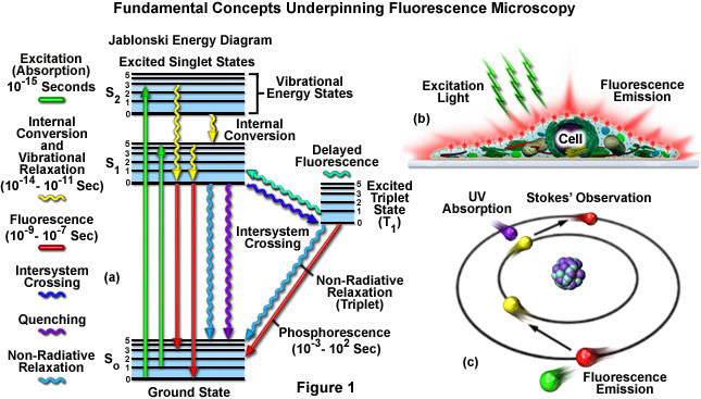
ZEISS Microscopy Online Campus, Microscopy Basics
Cell size homeostasis is maintained by CDK4-dependent activation of p38 MAPK - ScienceDirect
PDF) Nitrogen Stimulates the Growth of Subsurface Basalt
Chemically induced reprogramming to reverse cellular aging
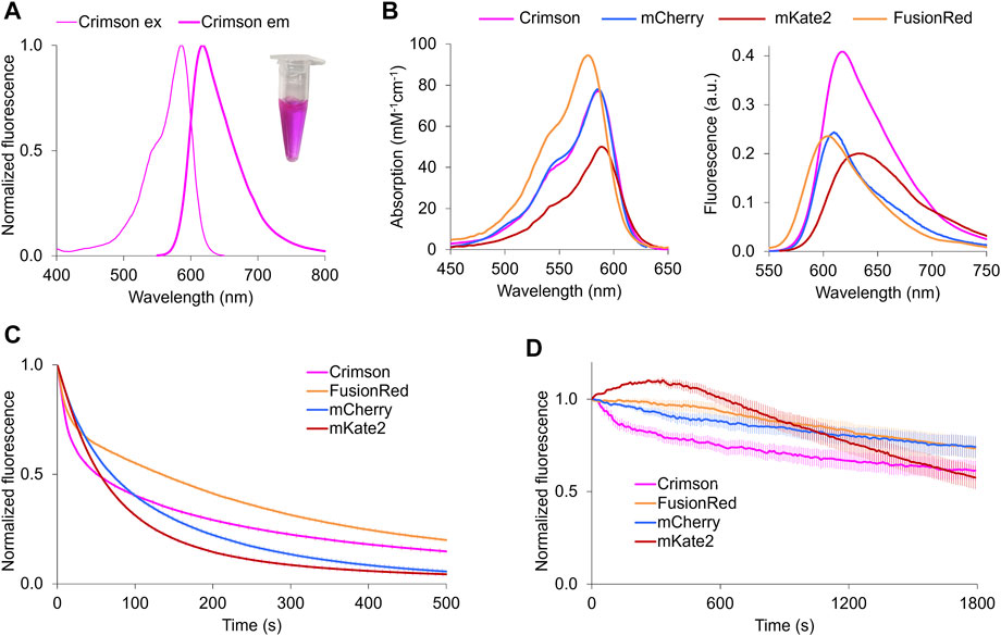
Frontiers A Bright, Nontoxic, and Non-aggregating red Fluorescent Protein for Long-Term Labeling of Fine Structures in Neurons
Cells, Free Full-Text
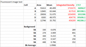
Measuring Fluorescence Intensity and Area – Keith R. Porter Imaging Facility – UMBC
Flow cytometry is a powerful tool for cell
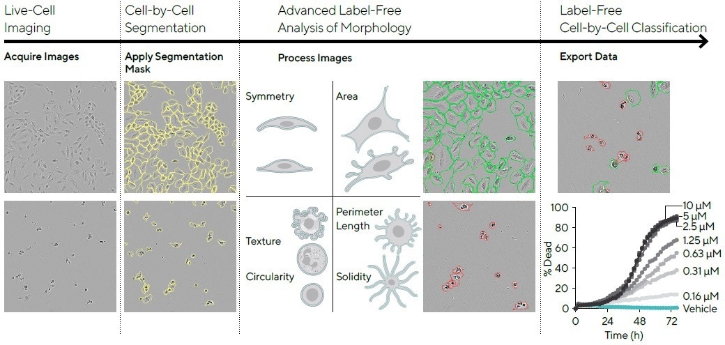
Cell morphology subpopulation classification in a label-free method